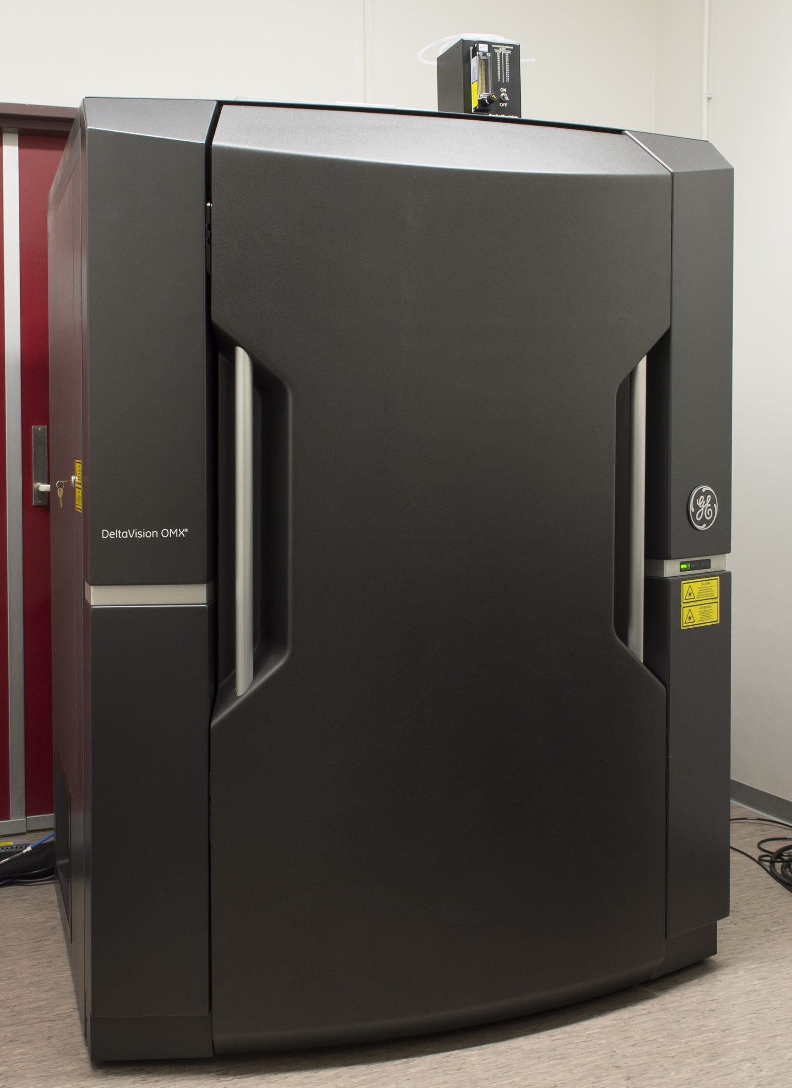
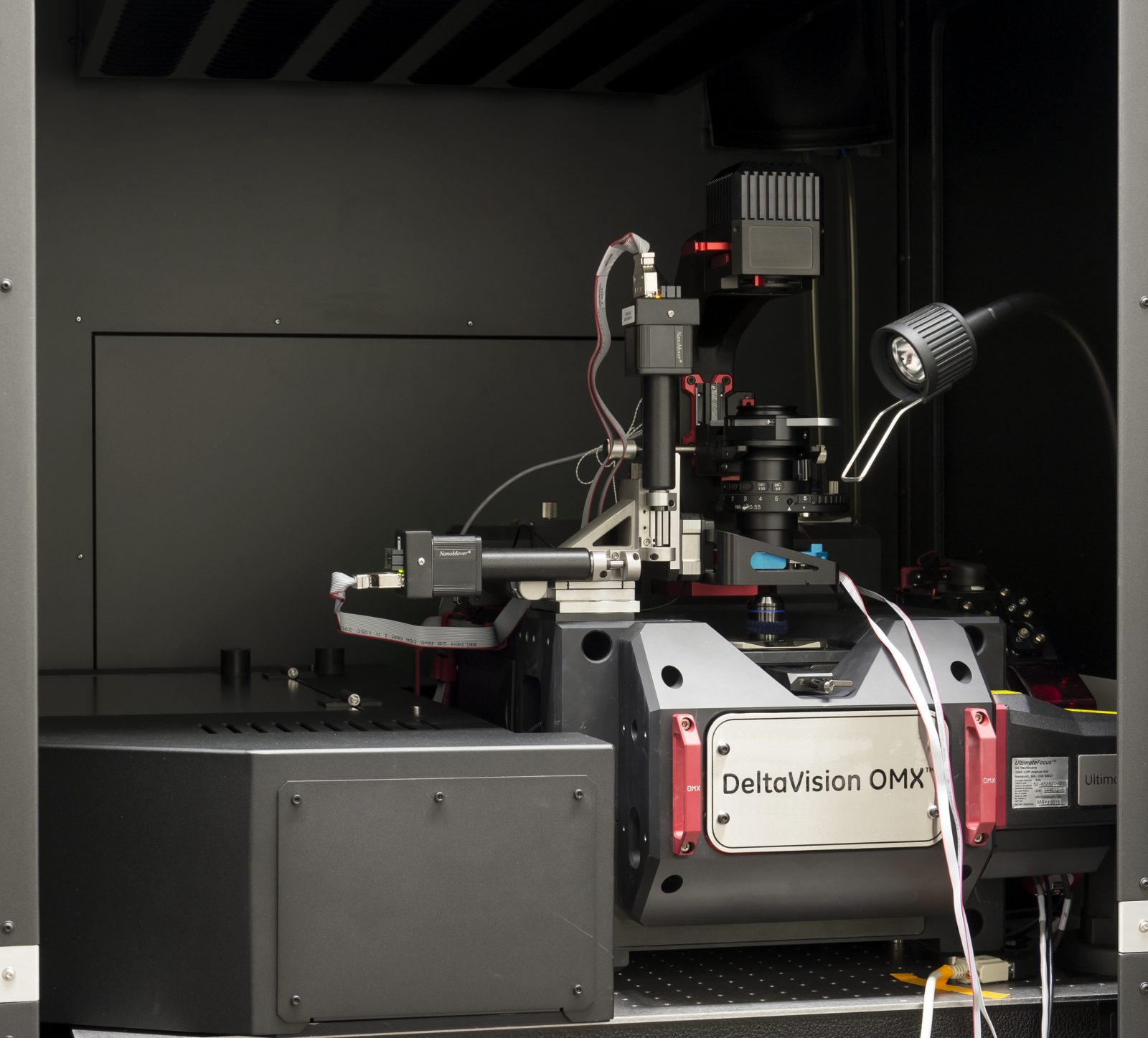
OMX-V4 Blaze


 Info about the system was on the GE-healthcare website we summarize the capabilities of the OMX-V4 here. GEH sold its life sciences branch to Danaher (which also covers Leica microsystems), the company is called Cytiva at this time.
An excellent website that displays imaging capabilities of the system is from the Harvard Medical School (dr. Waters’ imaging facility). There is a nice video outlining an identical setup at the Turku Bioimaging facility by one of the expert microscope engineers, Jeff Benz, in this link
Handouts:
Some advice on working procedures are on the desktop of the OMX master PC, we will keep these SOPs optimized for the setup when update or upgrades are implemented.
Quick reference:
- Startup and acquisition (PDF, yet you need to tweak settings)
- SIM deconvolution/reconstruction and registration (PDF).
On the Windows workstation desktop other documents are present for operating the system in detail.
Acquisition data from the Windows master PC are transferred automatically via the network circuitry to the LINUX workstation (in the CCI data folder). Acquisition and analyses can be performed in parallel because of this. Make sure that filenames do not have spaces, LINUX will not be able to process them.
NOTES:
– Realize that only the 25×25 mm central region of a slide can be imaged. The sample should be inside the central region, so only middle 4 wells of 8 well chamber can be visualized and coverslips with samples should centrally positioned on an object glass.
– When shutting down the system, be sure to place the stageholder to the top position as defined in the Z stage presets.
– an oil suggestion APP is available within your browser and Apple devices.
Special Add ons:
- Lasers:
- Diode 405 nm (Vortran Stradus, 100 mW)
- Diode 445 nm (Omicron LuxX, 100 mW)
- OPSL 488 nm (Coherent , 100 mW)
- OPSL 514 nm (Coherent , 100 mW)
- OPSL 568 nm (Coherent , 100 mW)
- Diode 640 nm (Vortran Stradus, 110 mW)
- LED illumination/ InSightSSI (Lumencor spectra X):
- 381-410 nm (104 mW)
- 426-450 nm (140 mW)
- 461-493 nm (110 mW)
- 505-520 nm (35 mW)
- 562-581 nm (125 mW)
- 638-653 nm (45 mW)
- Live cell imaging chamber (20-20/OKOlab -we are optimizing it)
- Hardware based Automated Focus control (Ultimate focus)
- 4 sCMOS (PCO) camera alignment for simultaneous image detection (CCD pixel size: 6.45 um, Image pixel size by 60x objective and 1.3 tube lens: 80 nm)
- PK module for fast Photoactivation procedures
- Pointillism/ SMLM based SuperResolution imaging module termed DLM, restricted for use in 2D
Speciality:
- Multicolor Live cell imaging (TIRF / Wide field )
- Deconvolution (SoftWoRx)
- SIM based super resolution microscopy
- DLM / pointillism / dSTORM- based microscopy (in 2D)
Available lenses:
- 60x TIRF lens (Olympus APO TIRFM , NA 1.49, WD=0.1 )
- 60x SIM lens (Olympus U-PLAN APO, NA 1.42, WD=0.15 )
Fluorescence Cube setup:
Drawer 1: Fluorescent Dye optimized (BGR)
| Arbitrary Name | Ex range | Em range | Suggested laser line |
| DAPI | 382-409 | 421-450 | 405 nm |
| FITC | 462-492 | 505-549 | 488 nm |
| Alx568 | 561-580 | 591-627 | 568 nm |
| Cy5 | 639-652 | 664-702 | 640 nm |
Drawer 2: Fluorescent Protein optimized (CYR)
| Arbitrary Name | Ex range | Em range | Suggested laser line |
| CFP | 382-409 | 421-450 | 445 nm |
| YFP | 462-492 | 505-549 | 514 nm |
| mCherry | 561-580 | 591-627 | 568 nm |
| Cy5 | 639-652 | 664-702 | 642 nm |

